Monday, January 11, 2010
INTRO FOR WEBLOGS

Monday, November 9, 2009
SPONDYLOSIS
 ·
·
Spondylosis is a condition of degenerative changes in the spine. Spondylosis can progress into spondylolisthesis. Spondylolisthesis is the slippage of one vertebra over another vertebra. This can occur in three different ways: forward (anterolistheis), backward (retrolistheis), or sideways (laterlistheis). Symptoms of spondylosis and spondylistotheisis are back pain, sciatica, numbness, difficulty walking, and burning sensations.
Monday, November 2, 2009
CLAY SHOVELER'S FRACTURE

A Clay Shoveler fracture is an uncommon avulsion fracture of the spinous processes, usually of c-7 or t-1. This fracture is caused by a sudden load on a flexed spine when the arms are extended. Shoveling heavy loads in rapid succession places the body in the perfect position for this to occur.
Clay Shoveler’s fracture causes a burning “knife” like pain in the upper back between the shoulder blades. The severity of the fracture determines treatment course. Often times no treatment is required and the fracture heals on its own. Pain medications and physical therapy are most common when treatment is necessary. In severe cases surgery is preformed to remove the tips of the spinous processes of the affect vertebra.
Tuesday, October 20, 2009
Jefferson Fracture
A
.
 c-1 fx
Plain radiographs, CT, and MRI are all used in the diagnosis of

CT image Jefferson Fracture
Monday, October 19, 2009
Carotid body tumors
Carotid body tumors are very uncommon. The tumor usually appears just lateral to top of hyoid bone at the upper part of the neck where the common carotids branch into the internal and external carotids. Carotid body tumors are also called chemodectomas or paragagliomas. They are slow growing tumor which is usually benign.
Carotid body tumors usually present with hoarseness and difficulty swallowing, but are mostly painless. They usually affect women over the age of 50. There are numerous modalities to detect carotid body tumors: CTA,
![[CBT2.jpg]](https://blogger.googleusercontent.com/img/b/R29vZ2xl/AVvXsEhaIfffc274kvwZ02oKdzMJ0BnIDgIUr2eus6OTHNVAImlQyYQPLFYu48NIrompaYJWzcHaso0_UtzL_IkMpQdwQN5KhFXueZ3qjC4_mcOFlJ81maEWhoRiKV1JEyU9yp_9-xE-kRNPGN-n/s1600/CBT2.jpg)
![[CBT4.jpg]](https://blogger.googleusercontent.com/img/b/R29vZ2xl/AVvXsEjvleaA0d9siA8b68DJn4O4fzCgiB-NmiAaithLDxotBoJGQJw1OI-I3-p-L6vTAC-E8YkeyPEYaZW_txuwwKuBNYdfLvZtez_rzHQLiZ4EqeLz2BYXMU006mECkh4U4blW1V81WZPxNT4k/s1600/CBT4.jpg)

Monday, October 12, 2009
Salivary Gland Calculus/Obstruction
Salivary glands produce saliva when we eat to help aid in chewing and digestion. Chemicals contained in saliva can crystallize to form a calculi or stone and block the ducts of the salivary glands. When a duct is clogged by calculi, this inhibits saliva from being able to exit the duct therefore causing pain and inflammation of the gland. The most common salivary glands to be affected by calculus are the submandibular glands, although any salivary gland can develop stones.
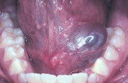
1 out of every 30,000 persons can be affected by salivary gland calculi. To determine if there is a stone or where it is located there are different modalities to choose from: CT, MRI, NM, and US.
 |
| stone
|
Usually the stone is crushed or removed via endoscopic procedure depending on the size and shape of calculi.
 |
|
|
|
|
| Salivary Gland Stone
|
Thursday, October 8, 2009
VEIN OF GALEN MALFORMATION
Vein of Galen malformation is a rare arterial venous malformation that develops between six and eleven weeks in utero. This causes an abnormal communication between the arteries and veins. Vein of Galen malformation typically occurs in new born babies with severe congestive heart failure, hydrocephalus, loud intracranial bruit, and seizure. An older child with a vein of Galen malformation will usually have an abnormally large head, developmental delays, learning disabilities, and strokes.
CT, MRI, and Angiography are all tools used to diagnosis vein of Galen Malformation. To repair this vessel, an embolization is preformed instead of traditional surgery. If this vessel is not repaired, it can lead to brain damage, stroke, or death.


