A
.
 c-1 fx
Plain radiographs, CT, and MRI are all used in the diagnosis of

CT image Jefferson Fracture
A
.
c-1 fx
Plain radiographs, CT, and MRI are all used in the diagnosis of
Carotid body tumors are very uncommon. The tumor usually appears just lateral to top of hyoid bone at the upper part of the neck where the common carotids branch into the internal and external carotids. Carotid body tumors are also called chemodectomas or paragagliomas. They are slow growing tumor which is usually benign.
Carotid body tumors usually present with hoarseness and difficulty swallowing, but are mostly painless. They usually affect women over the age of 50. There are numerous modalities to detect carotid body tumors: CTA,


Salivary glands produce saliva when we eat to help aid in chewing and digestion. Chemicals contained in saliva can crystallize to form a calculi or stone and block the ducts of the salivary glands. When a duct is clogged by calculi, this inhibits saliva from being able to exit the duct therefore causing pain and inflammation of the gland. The most common salivary glands to be affected by calculus are the submandibular glands, although any salivary gland can develop stones.
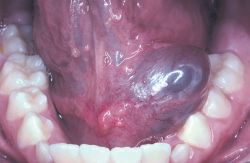
1 out of every 30,000 persons can be affected by salivary gland calculi. To determine if there is a stone or where it is located there are different modalities to choose from: CT, MRI, NM, and US.
 |
| stone
|
Usually the stone is crushed or removed via endoscopic procedure depending on the size and shape of calculi.
 |
|
|
|
|
| Salivary Gland Stone
|
Vein of Galen malformation is a rare arterial venous malformation that develops between six and eleven weeks in utero. This causes an abnormal communication between the arteries and veins. Vein of Galen malformation typically occurs in new born babies with severe congestive heart failure, hydrocephalus, loud intracranial bruit, and seizure. An older child with a vein of Galen malformation will usually have an abnormally large head, developmental delays, learning disabilities, and strokes.
CT, MRI, and Angiography are all tools used to diagnosis vein of Galen Malformation. To repair this vessel, an embolization is preformed instead of traditional surgery. If this vessel is not repaired, it can lead to brain damage, stroke, or death.


Angiofibromas are benign tumors of the fibrous connective tissue within the nasopharynx. They are invasive and aggressive. Angiofibromas are genetic and primary affect adolescent males.
Angiofibromas are known to cause tubercous sclerosis, which
is caused by an abundance of endothelium in the lined
vascular spaces.
Angiofibromas can cause nasal obstruction, abnormal speech, and difficulty swallowing.If left untreated, it can develop into ear canal obstruction.
Angiofibormas are visualized with either CT or MRI. There are
used to determine the size of the tumor and if there is any encroachment into other structures.
|
|
|
| ||||||
Angiofibromas are treated with surgery. Embolization of the tumor is preformed before the tumor is excised. If the tumor is not able to be removed, radiation may be utilized in an attempt to shrink the tumor’s size. |

Orbital cellulitis is an acute infection of the eye caused by a bacteria or fungus. It is sometimes a byproduct of injury, a sty, ethmoid sinusitis, or an insect bite. Orbital cellulitis usually affects only one eye. It can affect the globe of the eye, the upper cheek, the eyebrow, or even the eyelid. Most patients present with swelling or redness around the eye, high fever, and/or painful eye movement.
Time is very critical in the treatment of orbital cellulitis. If left untreated, it can lead to blindness, brain abscesses, or meningitis. When a patient presents with eye swelling, and fever, usually a CBC and blood cultures are drawn. A sinus x-ray and a contrasted CT are preformed in order to assist in the diagnosis.

The severity of the infection determines the treatment course. Generally, the patient is admitted to the hospital to receive IV antibiotics. If the IV antibiotics prove to be ineffective, sometimes it is necessary to go in and surgically decompress the infection.
Prolactinomas are benign tumors of the pituitary gland. They are the most common type of pituitary gland tumors. Prolactinomas can cause an increase or decrease the amount of the hormone prolactin. Changes in prolactin levels affect women and men differently. Prolactinomas are more common in women than in men and rarely affect children. Women may experience milk production (galacotorrhea), missed menses (amenorrhea) or irregular menses, and reduced sex drive (libido). Men may develop enlarged breasts (gynecomastia), low libido, vision loss, and headaches.
Blood tests are preformed to check for high levels of prolactin. MRI is the best imaging modality to determine if a tumor is a prolactinoma. Prolactinomas are usually too small to be visualized on CT.
 |  | |||
| Prolactinoma. | | Prolactinomas |
Prolactinomas are usually treated with medications that mimic Dopamine. Dostinex is on drug that is used in treatment of prolactinomas because of its dopamine like effect on the pituitary. Dopamine controls the amount of prolactin the pituitary gland produces. Medications usually control symptoms and patients are able to live a normal life. If the tumor has not shrunk or symptoms are not controlled by medication within six months of starting them, surgery or stereostatic radiation is recommended.


